A First Look at Your Smile
A first orthodontic visit is a chance to discuss what you would like to change and what questions you have. At Trielle Orthodontics in Union, NJ, that conversation supports an individualized assessment, whether you are exploring braces, clear aligners, or adult orthodontic care.
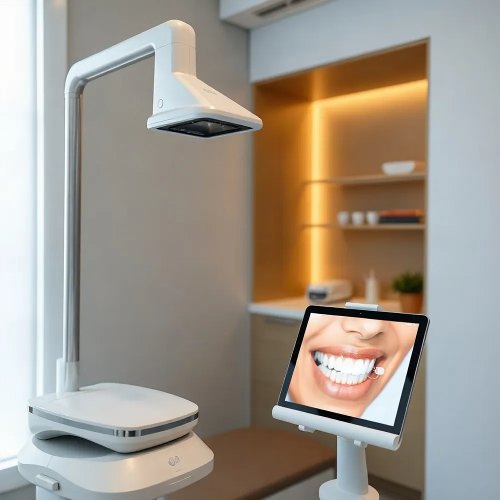
A digital smile scan can create a three-dimensional record of tooth surfaces and how the upper and lower teeth meet. The model gives the orthodontist a visual aid for discussing alignment and bite, but it does not show every structure beneath the gums. Intraoral scanners build these models from optical data.
The scan is one part of an assessment, not a treatment decision on its own. Your clinician may consider your concerns, goals, examination findings, and other records when discussing next steps. At Trielle Orthodontics, treatment planning is personalized, and the records recommended depend on the individual.
What a Digital Smile Scan Shows
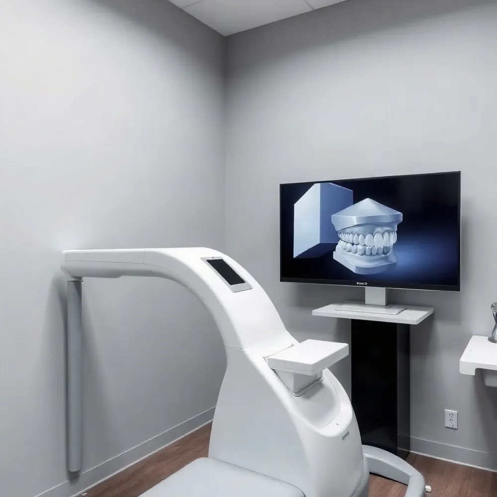 A digital dental impression is a three-dimensional model of the teeth and nearby oral structures, captured with a handheld intraoral scanner instead of impression putty. The scanner records surface details, and software assembles them into a model that can be viewed on a screen.
The model can show tooth position and spacing, along with how the upper and lower teeth meet. An orthodontist can rotate or enlarge the image to discuss areas of crowding, gaps, or bite alignment with the patient. This visual record can make findings easier to understand than a description alone. Intraoral scanners capture surface information, rather than showing structures hidden beneath the gums.
At Trielle Orthodontics, digital records can be considered alongside a patient's goals and clinical assessment when discussing individualized next steps. Later scans may also be compared with earlier records to help track changes in tooth position during care. The model supports assessment and planning, but it does not determine by itself whether treatment is appropriate.
Orthodontists may use digital models to plan custom appliances, including clear aligners and retainers, and to support planning for braces. The records and appliances recommended depend on the individual case. A scan is one part of the evaluation, not a promise that orthodontic treatment will be recommended or that a particular result is guaranteed.
What Happens During the Scan
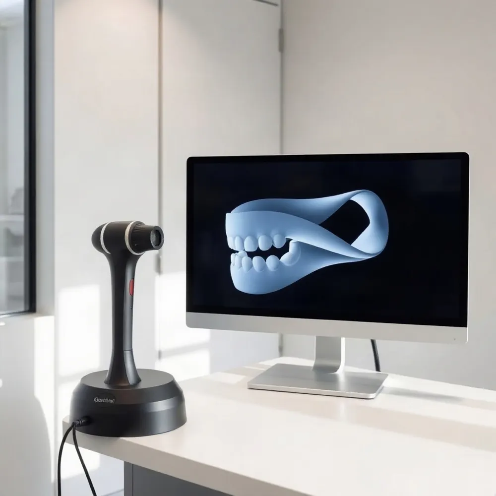 A clinician guides a small handheld wand over your teeth and gums. The scanner uses light to capture surface details, while software combines the images into a three-dimensional model. This process is known as intraoral scanning.
The model may appear on a nearby screen as the scan progresses. The clinician can check which areas have been captured and revisit spots that look incomplete. You may be invited to look at the model, though the clinician will explain what it can and cannot show.
During the scan, you may need to keep your mouth open briefly or shift your jaw so the wand can reach different surfaces. The process does not require a tray filled with impression material that sets in your mouth. The time varies depending on the areas being scanned and the clinic’s workflow, so a specific duration cannot be assumed.
If your jaw feels tired, an area is sensitive, or you need a pause, tell the clinician. The scan can be paused, and an incomplete area may be captured again if needed. Sharing concerns helps the team adjust the process to your comfort.
At Trielle Orthodontics, personalized care and patient education are part of the practice’s approach. Ask how the scan will inform your evaluation and what the clinician sees in the model. A professional evaluation can help connect those findings with your goals and possible next steps.
Accuracy and Scan Limitations
Digital impressions can capture detailed three-dimensional models that support many orthodontic tasks, including treatment planning and designing appliances. An intraoral scanner records surface data and software assembles it into a model, but the result is not automatically perfect for every patient or purpose. Intraoral scanners are one source of digital impression data, and their usefulness depends on the case and the quality of the capture.
Several factors can affect scan quality. The clinician’s scanning path and technique matter, as do access to tight spaces or the backs of teeth, saliva, reflections from surfaces, and movement of soft tissue. Processing software also affects how captured images are combined, so a clear view on screen does not by itself guarantee that every area has been recorded accurately.
The orthodontist can inspect the model as it develops, identify areas that look incomplete or unclear, and recapture them when needed. If a scan does not provide enough information for a particular clinical question, the provider may recommend another assessment or type of record. At Trielle Orthodontics, individualized treatment planning and patient education provide a basis for discussing what the scan shows and whether additional records are appropriate.
Patients can ask whether a digital impression is suitable for their specific treatment, which areas it captures, and whether any further records are needed. That conversation helps clarify both what the model can support and what it cannot establish on its own.
Digital and Conventional Impressions
A digital impression uses an intraoral scanner to capture tooth surfaces and build a three-dimensional model. A conventional impression uses a tray filled with material that sets around the teeth, creating a physical mold. Both methods can provide records for planning care, and the right choice depends on the clinical question and appliance being made.
Scanning avoids the taste and texture of impression material in the mouth. The model can be reviewed soon after capture, and the clinician can revisit areas that appear incomplete or need more detail. These features may make scanning easier for people who dislike impression trays, though comfort varies from person to person. Research comparing digital and conventional impressions describes the advantages and tradeoffs of each method in clinical use in a review of digital impressions.
Digital does not automatically mean faster or better in every case. The time required can vary with the patient’s mouth, the area being recorded, and the purpose of the impression. Saliva, limited access, or other scanning conditions can make some areas harder to capture, and the clinician may need to collect more detail or choose another method. An intraoral scanner records surfaces optically and produces a model for review using a digital scanning process.
Conventional impressions remain an option when they fit the clinical situation or the appliance being planned. Your orthodontist can weigh the available methods against the information needed for your care rather than treating one approach as suitable for everyone.
At Trielle Orthodontics in Union, NJ, care plans are individualized, with options that include braces and clear aligners. Ask the orthodontic team which impression method is appropriate for your assessment and planned treatment, and what to expect from that process.
When Additional Imaging Is Considered
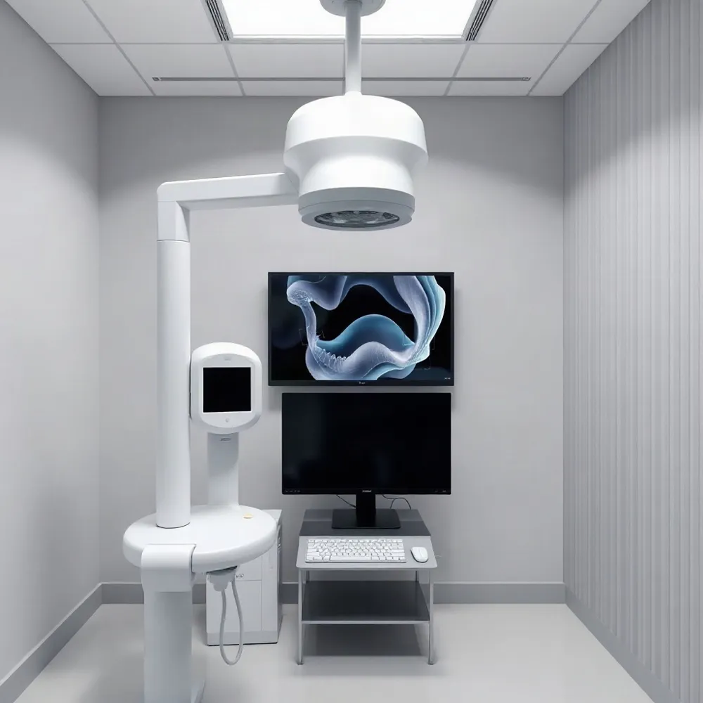 A digital surface scan and an X-ray provide different kinds of information. An intraoral scan records visible tooth and gum contours, while X-rays can show structures beneath the surface, such as roots and bone. The records answer different questions and are not interchangeable.
A three-dimensional intraoral model is also different from a cone-beam computed tomography (CBCT) scan. The intraoral scanner uses light to capture surface details; CBCT uses X-rays to create a three-dimensional view of teeth and jaw structures. A clinician may consider CBCT when the examination raises a specific question about, for example, an impacted tooth, root position, or jaw anatomy. CBCT imaging can provide information beyond what a surface scan shows.
Not every orthodontic patient needs CBCT or other advanced imaging. The records selected depend on the examination, the patient’s teeth and jaw, and the treatment question. For many patients, a digital impression alongside other appropriate assessment records may provide enough information to plan care. At Trielle Orthodontics, treatment plans are individualized, so the assessment should reflect the person’s needs rather than assume one set of images fits everyone.
Is a 3D dental scan necessary for orthodontic treatment? Not always. A surface scan can help document tooth positions and bite, but additional imaging may be considered if the clinician needs to assess structures that the scan cannot show. Patients can ask why a particular record is recommended, what it may clarify, and how the information could affect treatment decisions. Clear explanations help patients understand the purpose of each step without assuming that advanced imaging is routine.
Questions Make the Visit Clearer
A first scan is a way to collect information and talk through what it may mean. It does not obligate you to begin orthodontic treatment. At Trielle Orthodontics, care is based on an individualized assessment, with options such as braces or clear aligners considered according to a patient’s needs.
Ask the orthodontist to point out what the model shows, including any areas of crowding, spacing, or bite concerns. You can also ask whether additional records are needed, why they may help, and what choices could follow. An intraoral scanner creates a digital model from images captured inside the mouth, but the model is one part of an orthodontic evaluation, not a diagnosis on its own (intraoral scanner).
If anything is unclear, ask for an explanation before deciding on next steps. Understanding the findings and available options can make the visit more useful and help you choose what feels right for you.