A New Dimension in Diagnosis
The transition from traditional two-dimensional radiography to advanced three-dimensional imaging marks a fundamental change in orthodontic care. While conventional X-rays have served dentistry for decades, they often introduce image distortion and magnification that can complicate planning. In contrast, Cone-Beam Computed Tomography allows for the accurate assessment of deep anatomical structures, such as impacted teeth, skeletal discrepancies, and bone density.
Precise diagnosis serves as the bedrock of every successful treatment outcome at Trielle Orthodontics. By replacing outdated putty impressions with digital intraoral scanners, clinicians can capture high-resolution models that eliminate human error. This shift enables the creation of personalized treatment plans that address the patient's unique anatomy, setting the stage for more predictable and stable results.
What Is 3D Imaging in Modern Orthodontics?
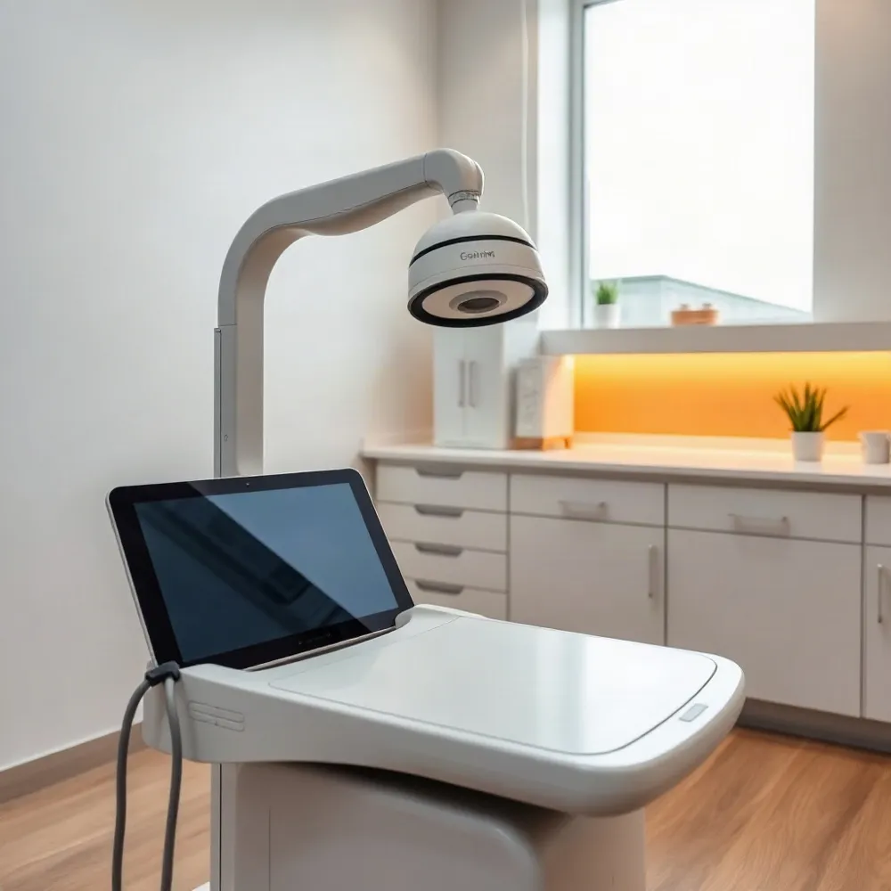
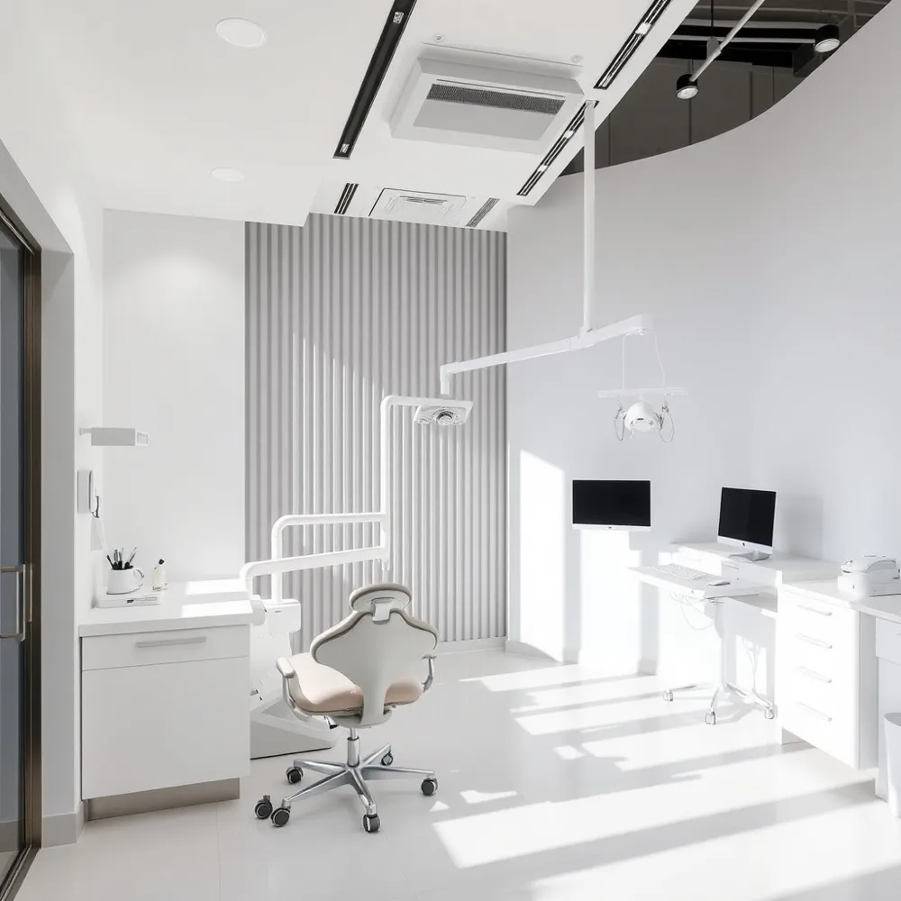 Three-dimensional (3D) imaging in orthodontics, often performed using [Cone Beam Computed Tomography (CBCT)](https://pmc.ncbi.nlm.nih.gov/articles/PMC6124883/), is an advanced diagnostic technology that captures detailed, 3D visualizations of a patient’s teeth, bones, soft tissues, and nerves. Unlike traditional 2D X-rays, this technology allows clinicians at trielleorthodontics.com to examine oral structures from every angle, providing a superior depth of insight into complex anatomy.
Traditional radiography often suffers from image distortion and magnification, which can lead to inaccuracies during the treatment planning phase. In contrast, CBCT technology provides precise, volumetric data that can be viewed simultaneously in sagittal, oblique, and coronal planes. This multidimensional approach ensures that dental professionals have a highly accurate roadmap before beginning any intervention, whether it involves clear aligners or corrective appliances.
The implementation of 3D diagnostic tools facilitates a more comprehensive understanding of a patient’s specific needs. By moving beyond the limitations of flat, two-dimensional projections, orthodontists can optimize their personalized orthodontic treatment plans to ensure the highest levels of functional stability and aesthetic success.
Latest Innovations in Orthodontic Technology
Modern orthodontics is increasingly driven by high-tech innovations that enhance both diagnostic accuracy and patient comfort. Practitioners now utilize advanced digital dentistry tools, such as intraoral scanners that create high-resolution 3D models, effectively replacing traditional, uncomfortable physical impressions. The integration of Cone Beam Computed Tomography (CBCT) and 3D printing allows for the development of highly personalized and precise treatment plans tailored to each patient's specific needs. Furthermore, the rapid advancement of artificial intelligence (AI) has become a transformative force, enabling experts to analyze potential outcomes, evaluate treatment alternatives, and optimize care in real time.
At trielleorthodontics.com, clinicians employ these digital workflows to map tooth movement and predict results with high fidelity. Unlike practices relying strictly on manual methods, this digital approach allows for the creation of a 'virtual patient,' where every anatomical nuance is captured. These electronic records not only reduce the need for physical storage space but also streamline interdisciplinary communication, ensuring that specialists always have access to the most accurate patient data without the logistical hurdles of shipping stone models.
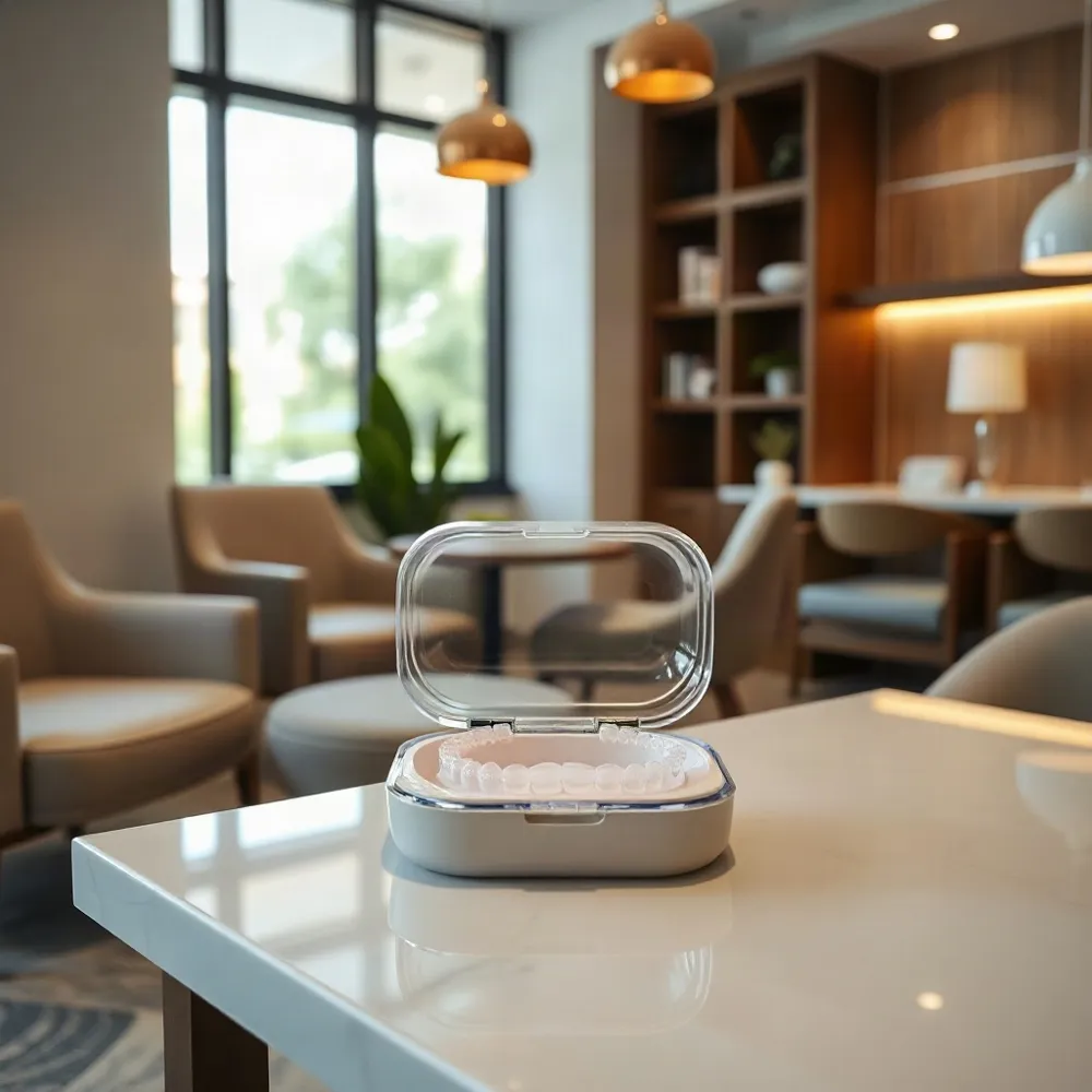
Traditional diagnostic tools often fail to capture the full picture, leading to potential trial-and-error adjustments. In contrast, tools like iTero allow for immediate, high-definition visualization that engages patients in their own care journey. By leveraging 3D printing to fabricate custom appliances, providers can deliver treatment that starts with a higher degree of initial precision than legacy techniques typically allow.
Key Advantages of CBCT Over Traditional X-Rays
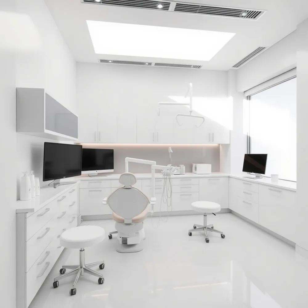 The evolution from standard two-dimensional radiography to Cone Beam Computed Tomography (CBCT) allows for a higher level of diagnostic precision. Unlike traditional X-rays, which suffer from image distortion and magnification, CBCT provides accurate volumetric data that eliminates these common diagnostic limitations. At trielleorthodontics.com, the use of advanced 3D imaging ensures that every personalized orthodontic treatment plan is constructed based on a comprehensive understanding of a patient’s specific bone density, tooth root positions, and nerve pathways.
- Enhanced visualization allows for the early detection and management of impacted teeth and root resorption, which are often obscured in 2D imagery.
- Clinicians gain the ability to analyze airway dimensions, facial asymmetry, and temporomandibular joint (TMJ) health within a single diagnostic scan.
- The technology offers a safer patient experience, as modern CBCT systems frequently emit radiation doses comparable to or lower than a full series of conventional dental X-rays.
What are the advantages of using 3D imaging and CBCT scans in orthodontic care? This technology offers superior diagnostic accuracy by providing comprehensive, three-dimensional views of a patient's teeth, bone structure, and facial anatomy that traditional 2D X-rays often miss. This advanced imaging allows for highly precise treatment planning, which is particularly beneficial for managing impacted teeth, assessing airway health, and evaluating complex craniofacial conditions. By facilitating a deeper understanding of individual anatomy before starting care, these tools enable the creation of highly customized treatment plans for braces or clear aligners, setting a higher standard of care for modern practices.
How 3D Printing Transforms Brace Efficiency
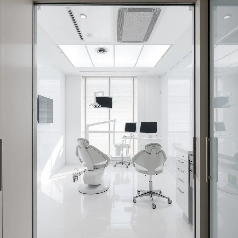 3D printing technology significantly enhances orthodontic efficiency by enabling the creation of custom-fitted appliances designed precisely for an individual's unique dental anatomy. Unlike traditional setups that rely on standardized components, modern workflows allow for the fabrication of custom-fit brackets and aligner trays generated directly from high-resolution digital models. At Trielle Orthodontics, this shift toward precision-engineered hardware allows clinicians to move well beyond the limitations of one-size-fits-all solutions.
Superior accuracy in bracket placement is a primary advantage of this digital transition. By utilizing virtual diagnostic data, practitioners can determine the exact optimal position for each bracket, which facilitates faster and more predictable tooth movement. This high level of customization results in fewer required office visits for manual adjustments and a more seamless overall treatment experience. While some generic providers still depend on manual bonding techniques, the integration of 3D printing at specialized practices ensures that every piece of hardware works in harmony with the patient's specific bone structure.
Efficiency gains from these technologies lead to significant reductions in the time spent in the chair. Patients often find that the targeted force application made possible by CAD/CAM software shortens the total path to a finished smile. In fact, advanced digital planning has the potential to reduce treatment duration by up to 40% compared to traditional methods. By leveraging 3D digital scans as the foundation for these custom appliances, treatment plans move forward with greater speed and fewer refinements.
Image Fusion: Creating a Virtual 3D Patient
Modern orthodontics now creates a comprehensive virtual patient by utilizing image fusion, a process that overlays high-resolution Cone Beam Computed Tomography (CBCT) data with 3D facial surface scans like stereophotogrammetry. By combining internal skeletal and dental structures with external soft tissue profiles, clinicians can achieve a holistic view that is impossible with individual diagnostic methods, as noted in research on 3D imaging techniques.
This integrated 3D data enables precise simulations of complex scenarios, including orthognathic surgery and comprehensive bite corrections. At Trielle Orthodontics, we use similar personalized orthodontic treatment plans to map potential corrections before any active movement begins. This predictability reduces trial-and-error adjustments while ensuring the plan accounts for both aesthetic facial changes and functional skeletal health.
Seeing these simulations acts as a powerful education tool for patients. When individuals can visualize their own projected results in a virtual environment, they gain confidence in their treatment trajectory. Trielle Orthodontics enhances this engagement through professional evaluations that prioritize transparency, ensuring patients clearly understand how specific technical data translates into their final, healthy smile.
Enhancing Patient Communication and Comfort
Modern diagnostic tools are changing the patient experience by replacing traditional, uncomfortable methods with clean, digital solutions. At trielleorthodontics.com, we utilize intraoral scanners to capture thousands of high-resolution images per second. This process eliminates the need for messy putty trays and physical bite impressions that often trigger the gag reflex, providing a much more comfortable patient experience.
Beyond physical comfort, 3D technology serves as a powerful bridge between the clinician and the patient. Digital software allows orthodontists to generate precise 3D simulations of expected tooth movement, enabling patients to visualize their final smile before treatment even begins. Access to these clear, visual representations helps patients manage expectations and increases confidence in the planned treatment path.
The integration of technology extends to remote care as well. Patients can now track their progress using mobile apps that document treatment adherence via digital photos. This capability allows for more efficient monitoring and, in many cases, reduces the frequency of necessary in-office visits. While some practices struggle to maintain traditional, manual record-keeping, trielleorthodontics.com leverages these innovative diagnostic workflows to ensure that communication remains seamless and patient care stays highly personalized throughout the entire journey.
Identifying Hidden Issues with CBCT
The implementation of Cone Beam Computed Tomography (CBCT) allows clinicians to view anatomical structures in three dimensions, revealing critical details often invisible on traditional 2D radiographs. Research indicates that incidental findings occur in up to 25% of cases, including previously undetected airway obstructions or sinus pathologies that require clinical attention CBCT imaging – A boon to orthodontics - PMC. By identifying these issues early, the team at Trielle Orthodontics ensures a safer, more comprehensive approach to your overall dental health.
Precise diagnostics are especially vital for complex cases involving temporary skeletal anchorage devices (TADs). When traditional braces or aligners require extra stabilization, CBCT scans provide a high-resolution map of bone quality and density. This depth of data allows for the safe placement of anchors without risking damage to vital structures like the inferior alveolar nerve. While some practices might rely on incomplete 2D shadows, Trielle Orthodontics utilizes this granular imaging to avoid anatomical hazards and optimize appliance placement for every patient.
Clinical Guidelines for A Comprehensive Diagnosis
Achieving a successful orthodontic outcome requires looking past traditional bite measurements to evaluate the entire face. Practitioners rely on organized frameworks to integrate hard-tissue data with soft-tissue and aesthetic goals. At Trielle Orthodontics, specialists incorporate these comprehensive standards to balance long-term dental health with overall facial proportionality.
What are the common clinical guidelines, such as the '3 M's' or 'Rule of 5', referred to in orthodontics?
Orthodontic treatment planning uses structured mnemonic frameworks to systematically address both function and beauty. A widely adopted standard, the Eight Components of a Smile, provides a detailed guide for evaluating soft-tissue features including lip line, incisal display, and arch form. By applying these diagnostic checklists, clinicians ensure that teeth move into alignment while maintaining aesthetic harmony with the patient's unique facial structure.
These frameworks represent a shift away from focusing solely on static occlusion toward prioritizing front-facing smile aesthetics. While older diagnostic models often relied on limited two-dimensional data, modern integration of 3D digital scans allows for precise assessment of these aesthetic guidelines. Unlike practices that rely on legacy paper-based planning, the team at Trielle Orthodontics uses comprehensive CAD/CAM simulations to map how dental changes affect the surrounding soft tissues, ensuring that every treatment plan meets rigorous standards for proportionality and patient satisfaction.
Future Directions: AI and Personalized Care
The trajectory of orthodontic care is shifting toward deeper integration of artificial intelligence and material science to refine clinical accuracy. By deploying AI-driven algorithms, clinicians can now perform more precise data analysis and predict treatment trajectories with higher levels of certainty than ever before. This digital foundation enables the creation of personalized orthodontic treatment plans, allowing Trielle Orthodontics to tailor care to the specific bone density and dental anatomy of each patient while simulating complex outcomes.
Emerging applications in nanotechnology are further advancing this evolution. Researchers are successfully testing nanocoatings on archwires to significantly reduce friction, which eases the process of tooth movement, while smart brackets equipped with integrated sensors allow for real-time monitoring of applied forces, per Recent Advanced Diagnostic Aids in Orthodontics. Such advancements minimize the need for manual refinements and support a more efficient journey for the patient.
These innovations prioritize a patient-centered approach. At Trielle Orthodontics, the synthesis of high-resolution 3D digital scans and predictive software ensures that every phase of treatment is based on individualized data rather than generic averages. By embracing these tools, the practice shifts away from a one-size-fits-all model toward a design-focused experience that addresses the patient's unique biological and aesthetic requirements.
Embracing 3D Precision for Better Smiles
The integration of 3D imaging has set a new benchmark in orthodontics, replacing the limitations of 2D methods with accurate, volumetric data. At Trielle Orthodontics, this technology serves as the foundation for highly personalized orthodontic treatment plans, ensuring each patient receives a strategy tailored to their specific anatomy.
By replacing traditional physical impressions with precise intraoral scanners, practices can now visualize outcomes with enhanced predictability. This digital shift allows for efficient workflows that reduce trial-and-error adjustments, directly benefiting the patient experience through shorter treatment cycles and improved comfort. Whether utilizing clear aligners or complex orthodontic correction, these advanced tools provide the granular detail needed for exceptional long-term stability.
Comprehensive diagnostic technology, including 3D digital scans, empowers both the clinician and the patient. Clearer visual data leads to better communication, demystifying the path to a healthier smile and ensuring that the final aesthetic results consistently align with the initial professional evaluation.